Microscopy Image Browser Features
Microscopy Image Browser is a high-performance software package for advanced image processing, segmentation and visualization of multidimentional (2D-4D) datasets. Microscopy Image Browser is written in MATLAB, but has a user friendly graphical interface that does not requre knowledge of MATLAB and can be used by a general audience without knowledge of programming.
Please find a list of main features below
Back to Index
Contents
-
List of all features -
List of Key Features: -
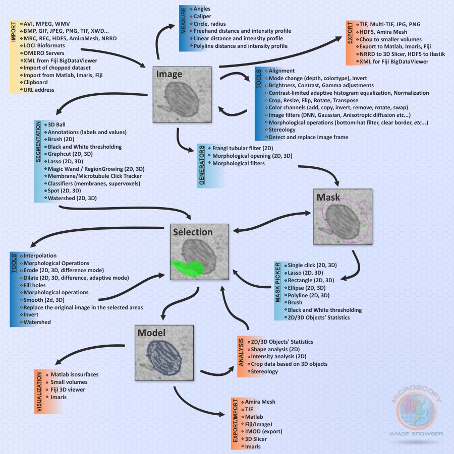
Scheme of Microscopy Image Browser
List of all features
Please check the list of all features page for more detailed description and video examples
List of Key Features:
- Works as a MATLAB program under Windows/Linux/MacOS MATLAB, or as a standalone application (Windows 64bit/MacOS/Linux)
- Open source licensed under GPLv3 license
- Compiled version is licensed for academic research
- Extendable with custom plugins (tutorials)
- Load/Import multiple image and video formats using standard and custom-made readers, Bio-Formats (LOCI) reader, OMERO server, and direct import from Fiji and Imaris
- Generation of multidimentional image stacks (3D/4D, width:height:color:depth:time)
- Batch processing mode for automatization of image processing steps
- Alignment of multi-dimensional stacks and images within these stacks
- Brightness, contrast, gamma, image mode adjustments, resize, crop functions, rotate, etc
- Semi-automatic/manual image segmentation with help of filters and interpolation in XY, XZ, or YZ planes
- Train and predict datasets using deep convolutional neural networks
- Quantification and statistics for 2D/3D objects
- Export of images or models to MATLAB, Amira , IMOD, STL, TIF, NRRD for 3D Slicer, Fiji BigDataViewer formats
- Direct 3D visualization using MATLAB isosurfaces and volume rendering, Fiji 3D viewer, and Imaris
- Filtering of images with core MATLAB and custom functions
- Log of performed actions
- Customizable Undo option
- Customizable Keyboard shortcuts
- Colorblind friendly default color modeling scheme
Scheme of Microscopy Image Browser
The connection diagram that illustrates the available tools and layers for image processing, segmentation, and visualization. This diagram showcases the workflow and interactions between different components in the software.
When working with opened images in Microscopy Image Browser, you have the flexibility to apply various filters and adjustments using a range of standard and custom functions. These functions enable you to enhance and modify the images according to your requirements.
Furthermore, MIB provides options for segmenting the images using variety of manual, semi-automatic and automatic techniques, including state of the art deep learning approaches.
The connection diagram below helps you understand the relationships between the different tools and layers, facilitating a seamless workflow for image processing and analysis.

Back to Index