Overview
Welcome to the home page of Microscopy Image Browser!
|
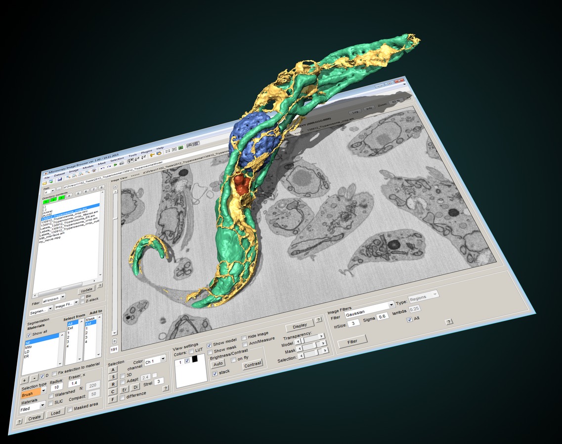
Microscopy Image Browser (MIB) is a high-performance MATLAB-based software package for advanced image processing, segmentation and visualization of multi-dimensional (2D-4D) light and electron microscopy datasets. |
Understanding the structure - function relationship of cells and cell organelles in their natural context requires multidimensional imaging. Advances in multimodal 3D imaging techniques have enabled a new insight into the morphology of tissues, cells and cell organelles that has not been conceivable before. As the access to such techniques is getting easier, effective processing, visualization, and analysis of a wide variety large datasets are posing a bottleneck for the research.
MIB is a freely available, user-friendly software for effective image processing of multidimensional datasets that improves and facilitates the full utilization of acquired data and enables quantitative analysis of morphological features. Its open-source environment enables fine tuning and possibility of adding new plug-ins to customize the program for specific needs of any research project.
Navigation
News and Updates
Actual versions
MIB MATLAB 2.93 [01.07.2026]
MIB Windows 2.93 [01.07.2026]
MIB Linux 2.93 [01.07.2026]
MIB Mac 2.91 [29.04.2025]
Update 2.91 highlights
 3D Segment-anything model 2
- New automatic alignment
- and more
Support
Please check list of all features for direct links to video examples
 Online call for help sessions are available on Fridays 15-16 Helsinki time
Online call for help sessions are available on Fridays 15-16 Helsinki time
Original publications
- Microscopy Image Browser: A platform for segmentation and analysis of multidimensional datasets
I. Belevich, M. Joensuu, D. Kumar, H. Vihinen and E. Jokitalo
PLoS Biology 2016 Jan 4;14(1):e1002340. doi: 10.1371/journal.pbio.1002340 - DeepMIB: User-friendly and open-source software for training of deep learning network for biological image segmentation
I. Belevich, and E. Jokitalo
PLoS Comput Biol. 2021 Mar 2;17(3):e1008374. doi: 10.1371/journal.pcbi.1008374