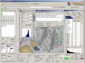
A screenshot of the MIB user interface. The program menu, toolbar, and panels are highlighted. A brief description of each element is provided.
A screenshot of the MIB user interface.
|
Main window of MIB. Additional panels responsible for image adjustments, objects quantification and action log are shown above the main window.
Main window and additional windows of MIB.
|
|
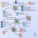
Connection diagram of available tools and layers. The opened images may be filtered and adjusted with number of standard/custom functions and further segmented either via the Mask route or directly with, for example, the brush tool and interpolation technique.
MIB organization diagram.
|
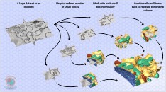
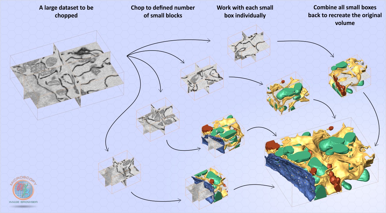
The large datasets can be chopped to smaller volumes to improve performance and do segmentation in parallel on several workstations. The chopped datasets and models can be automatically combined to reconstruct original volume
The chop mode
|
|
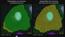
A special mode of the Brush tool in Microscopy Image Browser. This mode uses SLIC superpixels algorithm developed by Radhakrishna Achanta, Appu Shaji, Kevin Smith, Aurelien Lucchi, Pascal Fua, and Sabine Süsstrunk at EPFL (http://ivrl.epfl.ch/research/superpixels) for faster manual segmentation.
The Brush tool with SLIC superpixels.
Watch video
|
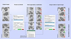
Rename and Shuffle workflow: the images are randomized and shuffled for classification or analysis, after which the models/classification results restored back
Rename and Shuffle
|
|
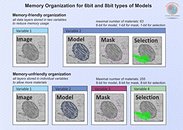
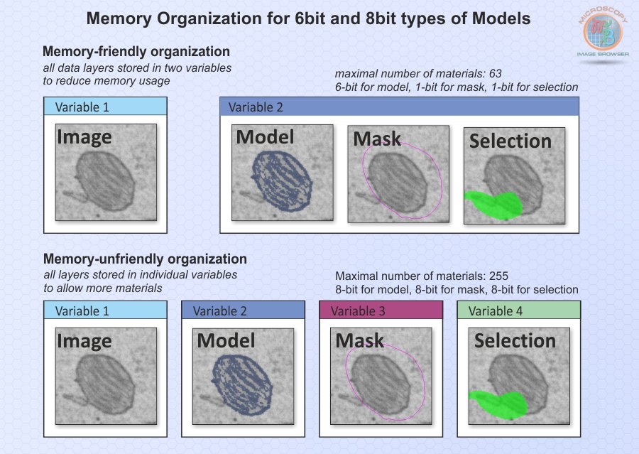
Memory organization of MIB. It is possible to use a single variable for keeping 3 service layers: Model, Selection and Mask reducing memory requirements.
Memory organization of MIB.
|
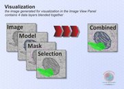
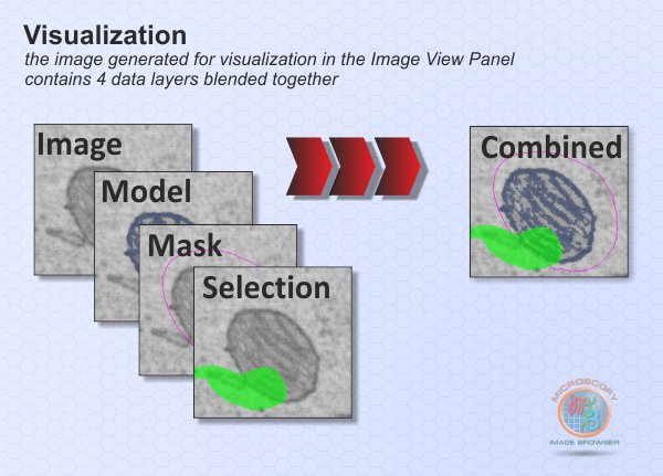
The final image that is shown in the Image View panel is composed of Image, Model, Mask and Selection layers blended together.
Blending of layers for visualization.
|
|

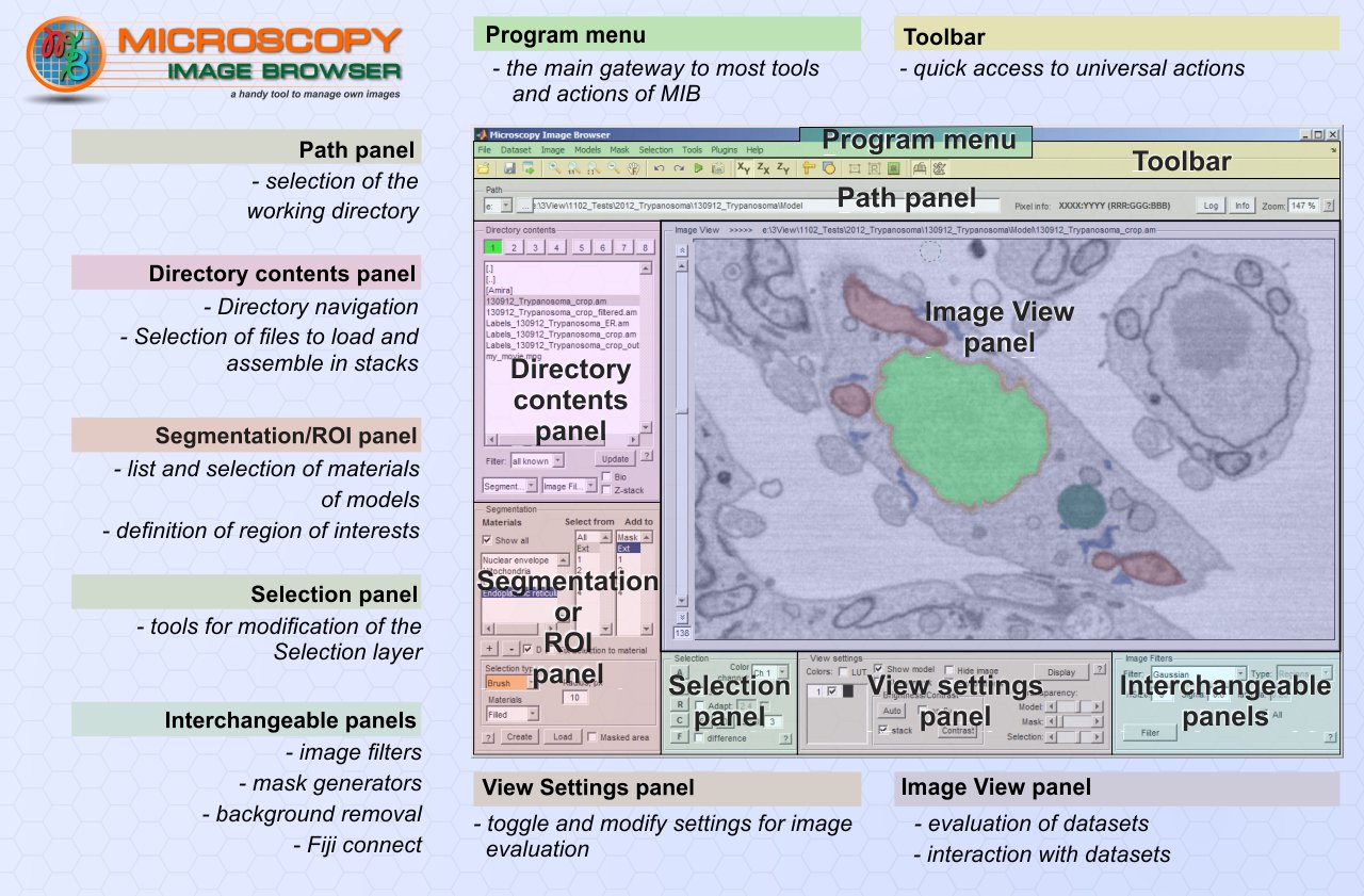 A screenshot of the MIB user interface.
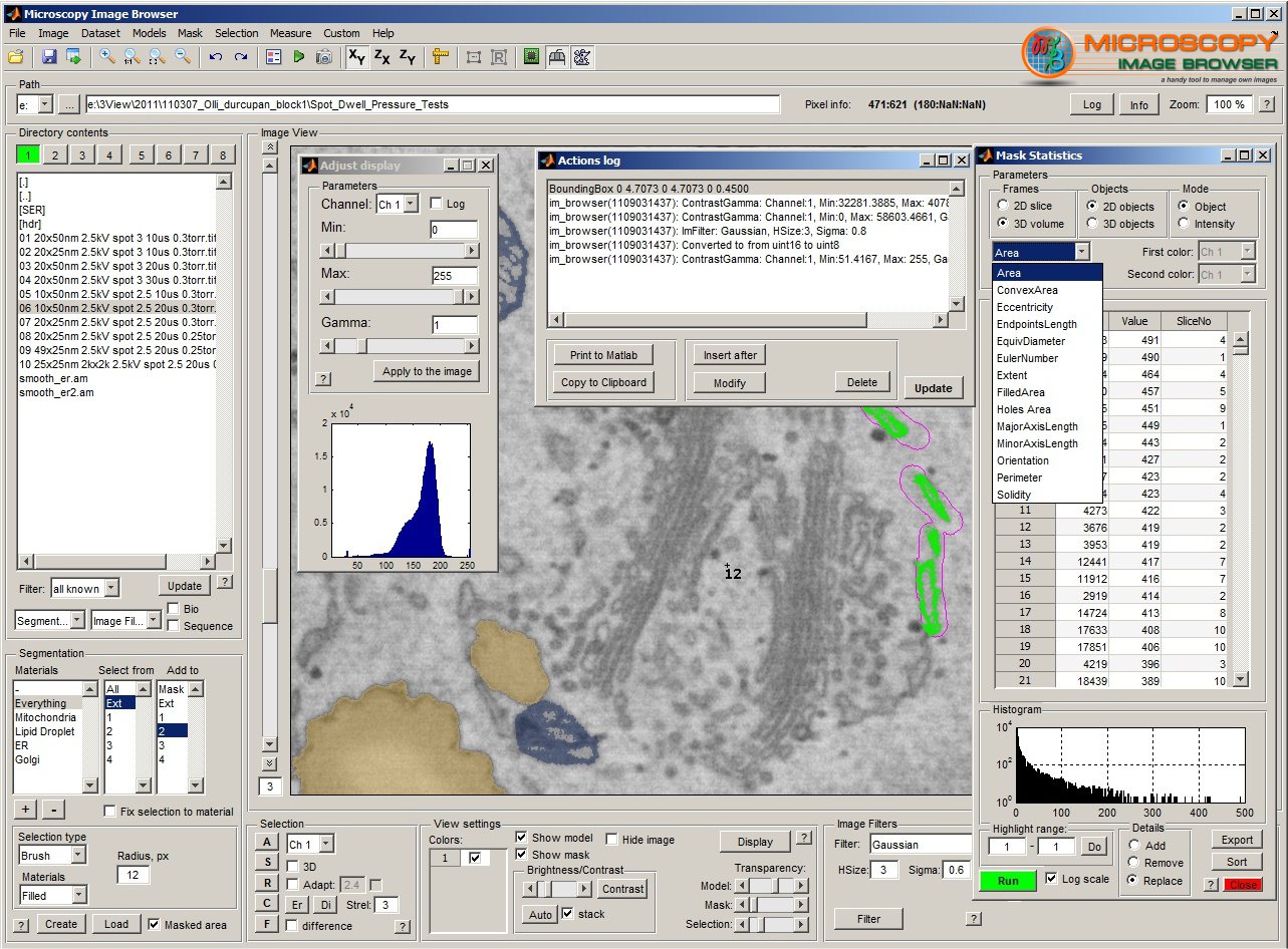 Main window and additional windows of MIB.
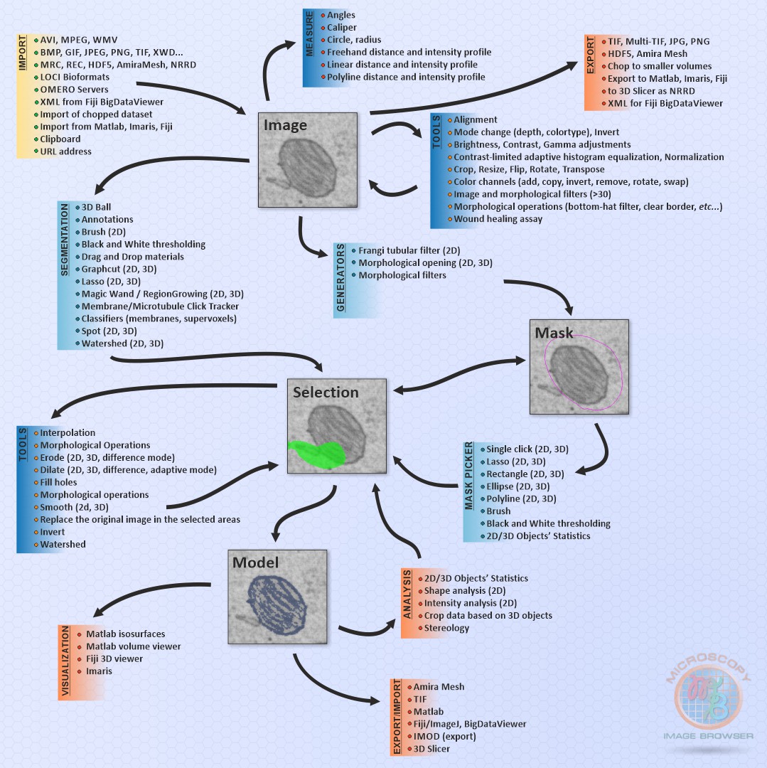 MIB organization diagram.
 The chop mode
 Rename and Shuffle
 Memory organization of MIB.
 Blending of layers for visualization.