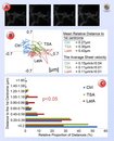
MIB was used to segment individual sheets of ER and analyse their dynamics using a custom made plugin.
A. Fragments of frames from a time-lapse video of human hepatoma cells (Huh-7/Hsp47-EGFP) with outlines of the segmented ER sheet. B. An example trajectories of Control, Latrunculin A (LatA) and Trichostatin A (TSA) treatments. C. Actin depolymerization (LatA) leads to significant (p<0.05) increase in the movement of centroids related to 1st centrome, while MT acetylation (TSA) has no effect on the movement.
Analysis of time-lapse video of control and treated cells obtained with light microscopy.
Joensuu M, Belevich I, Rämö O, Nevzorov I, Vihinen H, Puhka M, Witkos TM, Lowe M, Vartiainen MK, Jokitalo E. Mol Biol Cell. 2014 Apr;25(7):1111-26.
|
|


 Analysis of time-lapse video of control and treated cells obtained with light microscopy.
