File Menu
Provides access to some file handling actions for image dataset
Back to Index --> User Guide --> Menu
Contents
-
Import image from... -
Example datasets -
OMERO Import -
Batch processing... -
Chopped images... -
Rename and shuffle -
Export image to -
Save image as -
Make movie -
Make snapshot -
Render volume -
Preferences
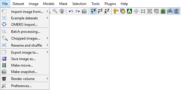
Import image from...
-
MATLAB - import image from the main MATLAB workspace into
MIB. It is possible to provide image description containers.Map together with the dataset, which allows to maintain parameters of the dataset when using theExport imagecommand (see below). [*brief demo*] - Clipboard - paste image from the system clipboard. This functionality is implemented using IMCLIPBOARD function by Jiro Doke, MathWorks, 2010. [*brief demo*]
- Imaris - import dataset from Imaris; requires Imaris and ImarisXT. This functionality is done with IceImarisConnector written by Aaron C. Ponti, ETH Zurich. [*demo*]
- URL - open image from provided URL address. The link should contain the protocol type (e.g., http://). [*brief demo*]
Example datasets
This menu entry provides quick access to several MIB demo datasets and full projects for image segmentation using deep learning. The datasets are grouped by techniques used for data collection.
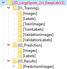
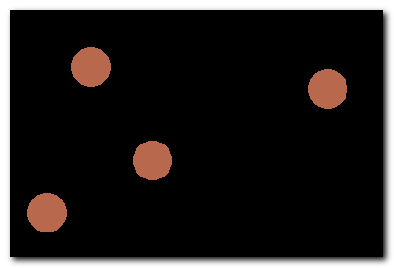
DeepMIB projects->Synthetic 2D large spots
The dataset includes a trained DeepLabV3-Resnet18 network for detection of large spots on a black background. The network can be opened by loading "2D_LargeSpots_2cl_DeepLabV3.mibCfg" file via
Menu->Tools->Deep learning segmentation->Options tab->Config files->Load
 |
 |

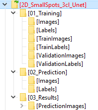
DeepMIB projects->Synthetic 2D small spots
The dataset includes a trained U-net network for detection of small random spots of 2 colors on a black background. The network can be opened by loading "2D_SmallSpots_3cl_Unet.mibCfg" file via
Menu->Tools->Deep learning segmentation->Options tab->Config files->Load
 |
 |

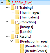
DeepMIB projects->Synthetic 2.5D large spots
The dataset includes a trained 2.5D DeepLabV3-Resnet18 and 2.5D U-net networks, where the 5-slice subvolumes were used for segmentation of large 3D spots on a black background. In addition, there are 2D spots that should not be segmented.
The trained networks are
- Spots_25D_DLv3RN18_Z2C_xy200z5 - DeepLabV3-based 2.5D Depth-to-Color network with patches of 200x200x5
- Spots_25D_Unet_Z2C_xy200z5 - U-net-based 2.5D Depth-to-Color network with patches of 200x200x5
- Menu->Tools->Deep learning segmentation->Options tab->Config files->Load
- Drag and drop of the config file into DeepMIB window
 |
 |

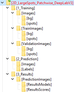
DeepMIB projects->Synthetic 2D patch-wise
The dataset includes a trained Resnet18 network for detection of patches that belong to large white spots on black background.
The network can be opened by loading "2D_LargeSpots_Patchwise_Resnet18.mibCfg" file via
Menu->Tools->Deep learning segmentation->Options tab->Config files->Load
 |
 |

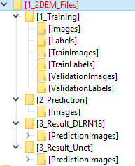
DeepMIB projects->2D EM membranes
The dataset includes a trained U-net and DeepLabV3-Resnet18 networks for semantic segmentation. This is an updated project that has been published with the DeepMIB paper: DeepMIB: User-friendly and open-source software for training of deep learning network for biological image segmentation, Figure 1a. Check readme.txt file for details.
The network can be opened by loading "2D_EM_membranes_Unet.mibCfg" or "2D_EM_membranes_DLabRN18.mibCfg" file via
Menu->Tools->Deep learning segmentation->Options tab->Config files->Load
 |
 |
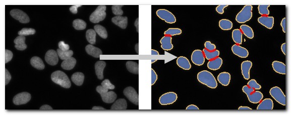
DeepMIB projects->2D LM nuclei
The dataset includes a trained U-net network for semantic segmentation. This is an updated project that has been published with the DeepMIB paper: DeepMIB: User-friendly and open-source software for training of deep learning network for biological image segmentation, Figure 1b. Check readme.txt file for details.
The network can be opened by loading "valid_252px_32patches_50ep.mibCfg" file via
Menu->Tools->Deep learning segmentation->Options tab->Config files->Load
 |
 |
DeepMIB projects->3D EM mitochondria
The dataset includes a trained 3D U-net network for semantic segmentation. This is an updated project that has been published with the DeepMIB paper: DeepMIB: User-friendly and open-source software for training of deep learning network for biological image segmentation, Figure 1c. Check readme.txt file for details.
The network can be opened by loading "NoValidation_valid_Aug_128px_256pat.mibCfg" file via
Menu->Tools->Deep learning segmentation->Options tab->Config files->Load
 |
 |
DeepMIB projects->3D LM inner hair cells
The dataset includes a trained 3D anisotropic U-net network for semantic segmentation. This is an updated project that has been published with the DeepMIB paper: DeepMIB: User-friendly and open-source software for training of deep learning network for biological image segmentation, Figure 1d. Check readme.txt file for details.
The network can be opened by loading "InnerEar3D_Hybrid_Same_136x64px_120ep.mibCfg" file via
Menu->Tools->Deep learning segmentation->Options tab->Config files->Load
 |
 |
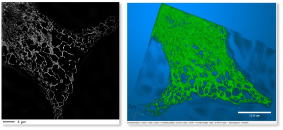
LM->3D SIM ER
A 3D super-resolution structured illumination light microscopy dataset of endoplasmic
reticulum
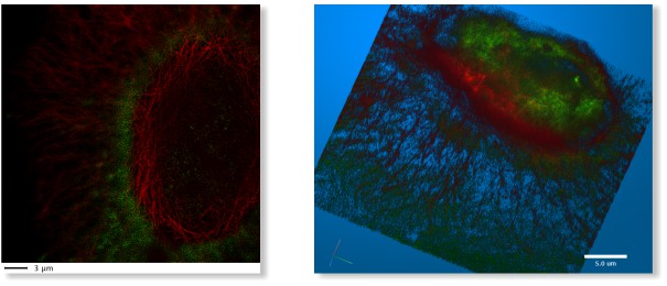
LM->3D STED
A 3D super-resolution Stimulated emission depletion (STED) microscopy dataset
LM->WF ER photobleaching
A wide-field time-lapse imaging of endoplasmic reticulum with visible
photo-bleaching effect

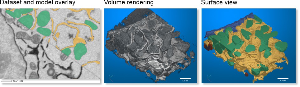
SBEM->Huh-7 and model
A small fragment of serial block face scanning electron microscopy
dataset featuring a Huh-7 cell and a model of nuclei, endoplasmic
reticulum, mitochondria and lipid droplets.
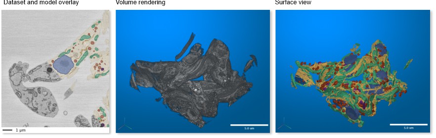
SBEM->Trypanosoma and model
A fragment of serial block face scanning electron microscopy
dataset featuring a cell of Trypanosoma brucei and a model of nuclei, endoplasmic
reticulum, mitochondria, vesicles, lipid droplets and cytoplasm.
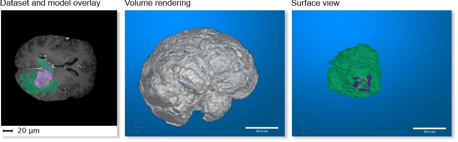
MRI->MATLAB Brain and model
One of test brain datasets from MATLAB taken with magnetic resonance
imaging (MRI) and a model of tumor. This dataset is only available in MIB
for MATLAB.
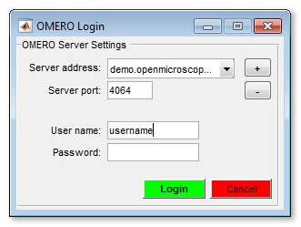
OMERO Import
Establish connection and load images from OMERO server. Requires OMERO server files to be downloaded. Please refer to the System Requirements pages for details of installation.
Selection of a server (please do not copy/paste the password!):
In the following dialog it is possible to select desired dataset and the range to take

A brief demonstration is available in the following video:
 https://youtu.be/iR7OL0eJGuw
Batch processing...
With the batch processing mode many of the image processing operations may be designed into an image processing workflow, which may automatically applied to multiple images.
Please refer to the Batch processing... section for details.
Chopped images...
This is a special mode that allows to split a large dataset into defined number of smaller ones and combine them back later. It is also possible to fuse the previously cropped dataset to the bigger one.
Please refer to the Chopped images... section for details.
Rename and shuffle
Two tools placed under this menu entry allow to shuffle files for their blind modeling, when the user does not know which file belong to which condition. The models from the shuffled files can be converted back over the original filenames for analysis.
Please refer to the Rename and shuffle section for details.
Export image to
Export images to the main MATLAB workspace or Imaris.
- MATLAB
In addition to the image
variable, another variable with parameters of the dataset (containers.Map class) is
automatically created. This |containers.Map| can be imported back to |MIB| later together
with the modified dataset to restore parameters of the dataset.
A brief demonstration on Import/Export is available in the following video:
https://youtu.be/zUJ1RUuTLVs
- Imaris
When Imaris is installed, the images can also be exported to Imaris.
Save image as
Save the open dataset to a disk.
The following image formats are implemented
- AM, Amira Mesh - as Amira Mesh binary format
- JPEG, Joint Photographic Experts Group - a method for saving lossy compressed RGB datasets
- HDF5, Hierarchical Data Format - saves images in the Hierarchical Data Format, version 5
- MRC, MRC format for IMOD - saves images in the MRC Format compatible with IMOD
- NRRD, Nearly Raw Raster Data - a data format compatible with 3D slicer
- OME-TIFF 5D (*.ome.tiff) - use BioFormats libraty to save the dataset as a 5D stack using OME-TIFF format
- PNG, Portable Network Graphics (*.png) - saves images in the Portable Network Graphics format
-
TIF format, LZW compressed, as a multilayered tif-file (
3D-Tif option) or as a sequence of tif-files (Sequence of 2D files). Please note! the TIF format uses 32-bit offsets, and that, in practice, limits the maximal size of the TIF-files to 2Gb. MATLAB can create TIF files larger than 2Gb but those can't be opened later - TIF format, non-compressed, see above for details of saving datasets in the TIF format
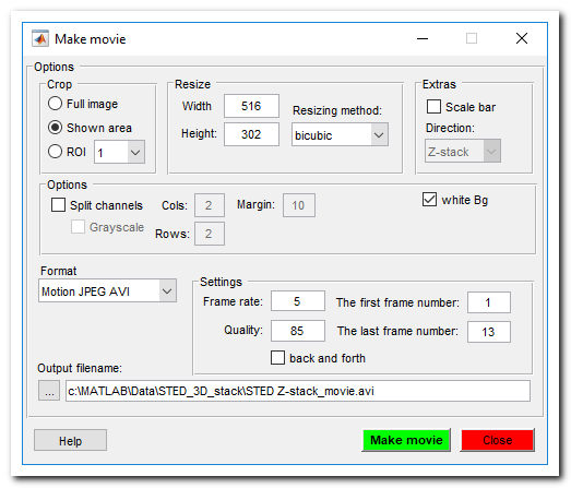
Make movie
Save dataset as a movie file. All objects that are shown in the image view window will be captured. Note! If the image width is too small the scale bar is not rendered. See more here.
Make snapshot
Make snapshot of the current slice. All objects that are shown in the image view window will be captured. See more here.

Render volume
3D visualization of volumes is available using 3 different ways
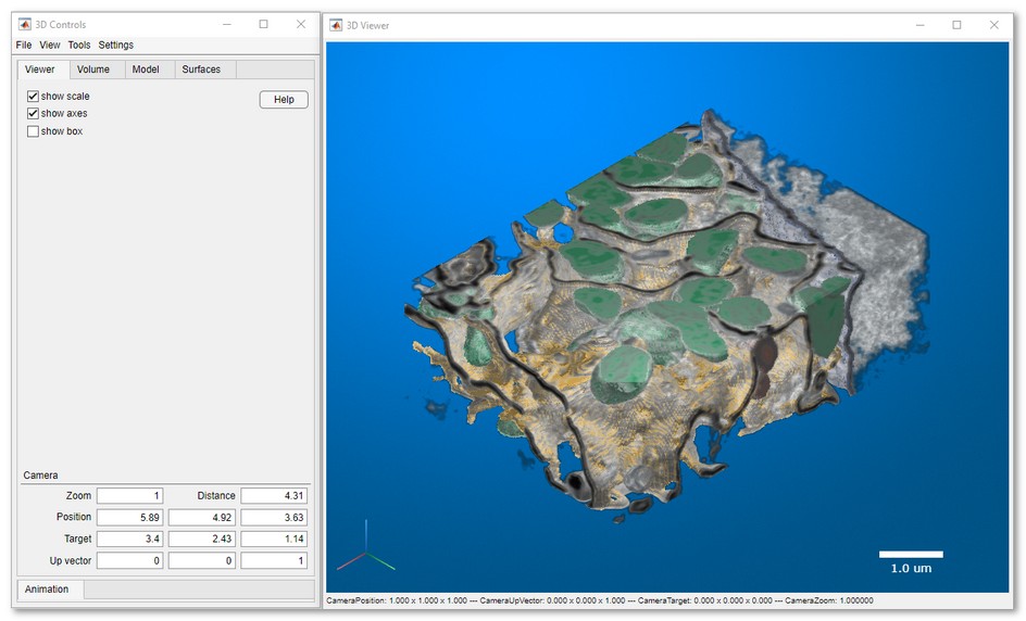
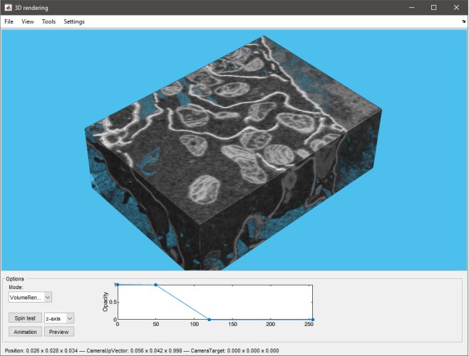
MIB rendering
Click to open documentation about the new 3D volume viewer is available
Starting from MIB (version 2.5) and MATLAB R2018b the volumes can be directly visualized in MIB using hardware accelerated volume rendering engine. The datasets for visuzalization can be downsampled during the export. It is possible to make snapshots and animations.
MIB version 2.84 comes with the updated 3D volume rendering that allows to render volumes with 1-3 color channels together with the models. As for MIB 2.84 the new 3D viewer is only available in MIB for MATLAB.
Volume rendering engines of MIB
| MIB 2.84, R2022b or newer
Volumes (1-3 colors) and models |
MIB 2.5, R2018b or newer
only 1-channel volumes or single material models |
 |
 |
Limitations
|
Limitations
|
The version of MIB rendering engine can be selected from preferences:
MIB->Menu->File->Preferences->User interface->3D rendering engine
A brief demonstration is available in the following video:
https://youtu.be/J70V33f7bas
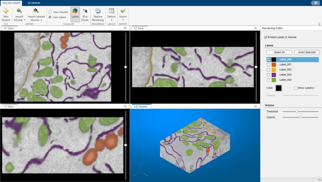
MATLAB Volume Viewer
When MIB is used with MATLAB R2017a or newer it is possible to export the open dataset to Volume Viewer application. Please note that this feature is not available for the compliled version of MIB
Snapshot of MATLAB volume renderer
A brief demonstration is available in the following video:
https://youtu.be/J70V33f7bas
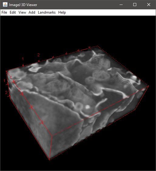
Fiji 3D viewer
Please refer to details in the Microscopy Image Browser System Requirements Fiji section for installation of Fiji.
Snapshot of volume renderering in Fiji
A brief demonstration is available in the following video:
https://youtu.be/DZ1Tj3Fh2HM
Additional dialog prompts for extra parameters for rendering:

- Reduce the volume down to, max width pixels - allows to reduce size of the dataset prior the rendering. This reduces memory consumption and improves performance. When this field contains 0 no volume resizing occurs.
- Smoothing 3d kernel, width - smooth the volume with gaussian blur. No smoothing when 0.
- Invert? [0-no, 1-yes] - invert the dataset, required for electron microscopy images.
-
Transparency threshold - all pixels with intensities below the provided value will appear transparent. Comma-separated numbers may be used here to provide specific transparency parameters for each color channel. In addition, the transparency threshold can also be tweaked in the Fiji 3D viewer window:
3D Viewer->Edit->Attributes->Adjust threshold.
Preferences
View and edit preferences of Microscopy Image Browser. Allows to modify colors of the Selection, Model and Mask layers, default behaviour of the mouse wheel and keys, settings of Undo. See more...
Please note, MIB stores its configuration parameters in a file that is automatically generated after closing of MIB
Location of the configuration file
-
for Windows - C:\Users\Username\MATLAB\mib.mat or in the Windows TEMP directory (C:\Users\User-name\AppData\Local\Temp\). The TEMP directory can be found and accessed with
Windows->Start button->%TEMP%command - for Linux - /home/username/Matlab or local TEMP directory
- for MacOS - /Users/username/Matlab or local TEMP directory
Back to Index --> User Guide --> Menu