Wound Healing Assay
Back to MIB | User interface | Menu | Tools
The wound healing assay is a microscopy-based technique used to study cell migration. A "wound" or gap is created in a cell monolayer, and the movement of cells into this gap is monitored over time using time-lapse imaging to assess healing dynamics.
Measures cell migration parameters, tested on the Imagen Cell-IQ platform with 0-pixel overlap image grids.
The tool has two parts: grid stitching of time-lapse datasets and the wound healing analysis
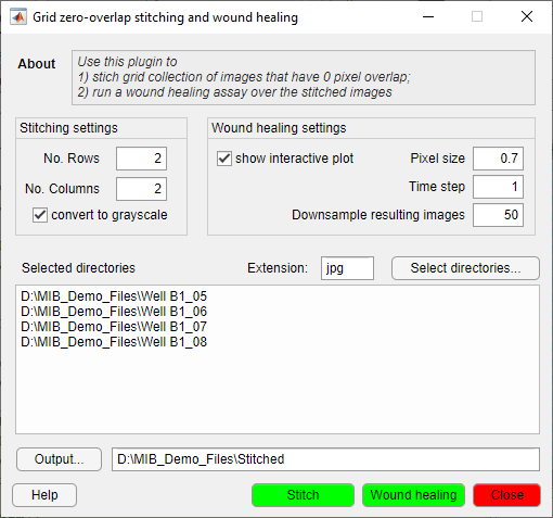
Stitching
- Use the Stitching settings panel to specify grid cell count (each cell in its own directory, named sequentially from top-left to bottom-right horizontally).
- Set filename extension.
- Select directories with grid images ().
- Specify output directory ().
- Start with the button.
Wound healing analysis
- Configure the Wound healing settings panel: pixel size, time step, and optional downsampling. Check for an interactive plot after each time point.
- Select directories with stitched images ().
- Start with the button.
- Plot of minimal, average, and maximal wound width.
- Excel sheet and MATLAB file with wound width values.
- Directory with wound snapshots.
- Text file with timestamps from original images.
Reference
Based on Cell Migration in Scratch Wound Assays by Constantino Carlos Reyes-Aldasoro.
Cite as: CC Reyes-Aldasoro, D Biram, GM Tozer, C Kanthou, Electronics Letters 44 (13), 791-793.
Code available on GitHub.
Back to MIB | User interface | Menu | Tools